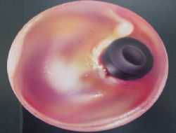
The middle ear is located behind the eardrum. It contains the bones of anvil, stirrups and hammer, which provide hearing.The middle ear is located behind the eardrum. It contains the bones of anvil, stirrups and hammer, which provide hearing. The sound coming from the external environment vibrates the membrane and is delivered to the auditory nerve and brain through these ossicles. The middle ear is in contact with the back of the nose (nasal) through the Eustachian tube. Thanks to the Eustachian tube, the middle ear receives air, and thus its internal pressure becomes equal to atmospheric pressure. If there is a dysfunction in the Eustachian tube, both the middle ear cannot air out and there is a collapse in the membrane due to negative pressure. Fluid accumulation begins insidiously inside the middle ear. The consistency and amount of this liquid gradually increases, initiating hearing loss.
In What Cases Can There Be Dysfunction in the Eustachian Tube?
In children, infectious agents from the nose and throat come to the ear more easily, both because of its narrow diameter and because it is in a more horizontal position. Past infections of this type can lead to adhesions and working disorders in eustachia. In addition, in enlarged nasal flesh and allergic noses, the Eustachian mouth is blocked by swelling of the surrounding tissues, leading to dysfunction. The presence of a congenital cleft palate is another reason for insufficient Eustachian function.
What is Ear Tube Surgery, When Should It Be Done?
It is necessary to equalize the middle ear pressure with atmospheric pressure. For this reason, a small window is needed on the eardrum. After a 2mm incision is made in the eardrum under general anesthesia, a silicone ear tube smaller than a matchstick head is placed on top of the membrane under a microscope image. The shape of the tube resembles a spool of thread. One end is behind the membrane, one end is outside. The operation takes about 20-30 minutes. It is necessary to insert a tube for recurrent serous otitis media (fluid in the middle ear) that does not heal with medication. During drug therapy, middle ear pressure is regularly monitored. Nasal flesh surgery is also performed in the same session in the vast majority of child patients. Thus, the Eustachian mouth will also be opened. In children without nasal flesh, the method of delivering compressed air through the nose before tube administration can be tried, but its success is limited. It is also useful to insert a ventilation tube into the eardrum for frequent recurrent middle ear infections (acute otitis).
How and When does the Tube Come Out? What Should I Pay Attention to After Surgery?
Although it depends on the patient, under normal conditions, it is enough for a tube to have worked healthily for 9 months. around 12 months, the tubes are spontaneously excreted into the external ear canal. If he didn’t throw it out on his own, his doctor decides when he should come out. Especially in recurrent diseases and in cases where repeated surgery is required, it may be desirable not to discard the tubes for many years. Entering the sea into the pool after surgery creates a concern that water escapes from the ear. The benefit of protection so that water does not escape from the outside of the ear is debatable.

