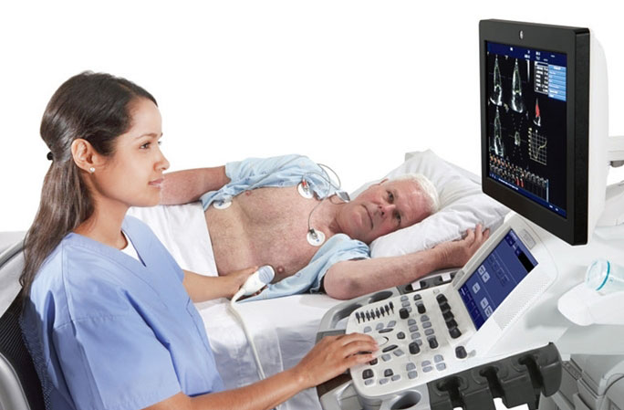
What is Echocardiography?
In echocardiography (also called echo for short), ultrasound (transonic waves) is used. Ultrasound waves are a sound beyond the hearing limit of the human ear (18,000 – 20,000 vibrations/sec). These sound waves are sent to the heart with the help of a sensitive instrument that is held by hand in the form of a tube and creates a sound that is passed around the patient’s chest. Sound waves return to the instrument from the heart walls, November muscles, valves. Different textures reflect sound waves in different ways. Thus, the sound waves returning from the heart are converted into a picture by a computer, and these images are printed on paper if desired, as they are viewed from a monitor. Echo is a quick and harmless test that provides important information about the heart.
With echo;
Heart valve diseases,
The diameters of the heart cavities, whether they are large,
Movements of the heart walls, whether there is a movement disorder (in cardiovascular diseases, wall movement disorders may occur where the relevant vessel is bleeding),
Intra-cardiac pressures,
Investigation of clots (thrombus) in the heart cavities,
The amount and percentage of blood that the heart beats at one time during contraction (After the human heart fills with blood, it throws out a certain percentage of the blood in it with contraction. The percentage of blood that the heart beats into the veins with each heartbeat is called the “ejection fraction”. The normal is around 55-70%. In other words, the heart can throw out 55-70% of the blood that comes to itself at one time.),can be investigated. In short, valuable information is obtained with echo on many topics such as cardiac rheumatism, valve diseases, heart failure, heart attack, congenital heart diseases.
Advantages of the economy:
It gives important information especially about the valve and wall movements of the heart,
There are many places,
Paint, radioactive material, needle are not used,
It is painless and not harmful to the patient.
The disadvantages of Eko are; The ability of the person performing the test affects the degree of accuracy of the test results. In some people, echo images are not good due to the person’s structure (the person is not echogenic) and sufficient images may not be obtained. Some parts of the heart may not look so good.
In addition, information about the coronary heart vessels and the viability of the heart muscle can also be obtained with the Stress ECHO performed with medication. Nov. With color Doppler echocardiography, information about the pressures of the heart cavities, the degrees of valve insufficiencies (leaks) are obtained.
In some cases, it may be necessary to obtain more detailed information via transesophageal (through the esophagus) echo (abbreviated as TEE). In this examination, the throat is numbed and a pinky finger-thick hose is inserted into the esophagus and the heart is examined from the back side and in closer detail. One should stay hungry before the TEE examination.

