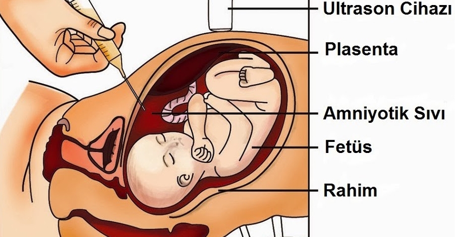
What is amniocentesis?
Amniocentesis is an operation to remove amniotic fluid from the mother’s womb with the help of a thin and long needle. The amniocentesis operation is usually performed between the 16th and 22nd of pregnancy. it is performed in risky pregnancies during the weeks. When performed by experienced doctors, it is quite painless and has few risks for the baby.
How is Amniocentesis Performed?
First, after a detailed ultrasound is performed on the expectant mother and the baby’s map in the uterus is removed, the skin of the abdominal area is wiped with an antiseptic substance. With a needle that is thin enough to be suitable for the procedure accompanied by ultrasound (usually needles used in spinal anesthesia are preferred), it is entered from a place in the abdomen that is suitable for the procedure, first into the uterus and from there into the uterine cavity where the amniotic fluid is located. The first 0.5 milliliter of the amniotic fluid drawn through the injector is excreted and a sufficient amount of fluid is withdrawn. After an ultrasound evaluation is performed again, the needle is removed from the place and the applied procedure is terminated. The received amniotic fluid material is sent to the laboratory provided that it is at room temperature. An average of 20 milliliters of fluid is taken in amniocentesis for genetic purposes.(up to 30 cc) This amount corresponds to about 10% of the total amount of amniotic fluid of a baby who is still at the 16th week of pregnancy. It is estimated that the baby completely replaces this liquid taken within a 3-hour period.
When Should Amniocentesis Be Performed?
Amniocentesis is currently most often used to accurately diagnose chromosomal abnormalities and other anomalies. The only difference between the applications made for treatment from a technical point of view is that they can be applied easily and easily during any period of pregnancy. Diagnostic amniocentesis is usually 16. it is applied from the week.
What is amniotic fluid?
Amniotic fluid, which is vital for babies, helps the baby to maintain its development in a pouch during pregnancy. This amniotic fluid is extremely important for the development of the baby throughout the entire pregnancy. The amnion protects the baby from external factors, helps the development of November muscles and nerves during pregnancy. Although the amount of amniotic fluid is not constantly constant, it is constantly absorbed and reproduced.
The main source of amniotic fluid is the baby’s lungs and kidneys. The baby swallows these liquids and passes into the maternal circulation with the help of the placenta. On the other hand, the urine that the baby extracts is an important source of amniotic fluid.
What is the source of amniotic fluid?
While the source of amniotic fluid is formed in the mother’s circulatory system during the first weeks of pregnancy 14. after the week of pregnancy, the baby’s urine is the most important source. The urine contained in the baby does not contain harmful waste substances, it is only an indicator of the filtration function of the kidneys. As the birth approaches, the proportion of fetal urine in the amniotic fluid gradually increases.
How is amniotic fluid measured?
In order to measure amniotic fluid, a concept called AFI (amniotic fluid index) has been developed. According to the weeks of pregnancy, there are also normal limits set for AFI. Thanks to this test, the level is determined. If it is above the limits, that is, if there is too much water, we call it Polyhydramnios, and if it is below it, there is a lack of amniotic fluid, that is, Oligohydramnios.
The presence of any of these conditions is not healthy at all for both the baby and the mother.

