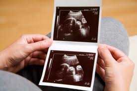
Structural anomalies can be seen in 2% or 3% of births. If it is necessary to open the content of anomalies, extremely simple ones and mental or structural disorders may have been observed. Genetic and environmental factors are the main factors in the structural disorders of the baby. After some severe anomalies observed in babies, the baby may be lost in the womb or immediately after birth before meeting with life. But against this, 2. level ultrasound is an important diagnostic tool. Some severe anomalies, such as Down syndrome, can continue to live. It can be treated with professional training and family support.
Early Diagnosis of Structural and Cormosomal Disorders by Ultrasonography
Ultrasonography is the most important tool for detecting structural disorders of the baby in the womb. The technology developed in ultrasonography in recent years and the improvement of image quality have greatly increased and sensitized the success rate of diagnosis. It has become possible to diagnose 60% of structural anomalies, especially with ultrasonography alone. As the rate of structural disorders in the baby decreases, the diagnosis rate may also decrease.
- What is Level Ultrasonography?
18 – 23. it is performed Decemberly between weeks. The general development of the baby and its organs are sufficiently developed for it to provide information on ultrasonography. The amount of water around the baby when taking an image is a favorable amount. In addition, there is still enough time for termination of pregnancy in case of severe structural impairment. In a detailed ultrasound, the structure of the mother’s uterus, the placenta, which we call the baby wife, and the baby itself are carefully examined by an obstetrician. With the measurements made, the growth tracking of the baby is calculated. The baby is here,
Central nervous system
Heart and chest cavity
Stomach and intestinal system
Kidney and urinary tract
Skeletal system
it is comprehensively monitored. All findings are noted and progress is calculated. The importance of second-level ultrasonography allows the physician to follow the birth process with appropriate treatment by taking precautions in advance for treatable anomalies.
11 and 14. Ultrasonography Performed During the Week Dec
In the last few years, some symptoms that have appeared during pregnancy and baby development have made the environment sufficient for ultrasonography between 11 and 14 weeks Dec. Thanks to the advanced ultrasonography devices also available in our hospital, it is possible to perform anatomical scanning basically even if the baby has not grown much in the womb. In babies known among the public, the measurement of the thickness of the nape can be seen by ultrasound performed between these weeks. Decapitation is a Decapitation procedure.

